
Foot Description, Drawings, Bones, & Facts Britannica
It is made up of over 100 moving parts - bones, muscles, tendons, and ligaments designed to allow the foot to balance the body's weight on just two legs and support such diverse actions as running, jumping, climbing, and walking. Because they are so complicated, human feet can be especially prone to injury.
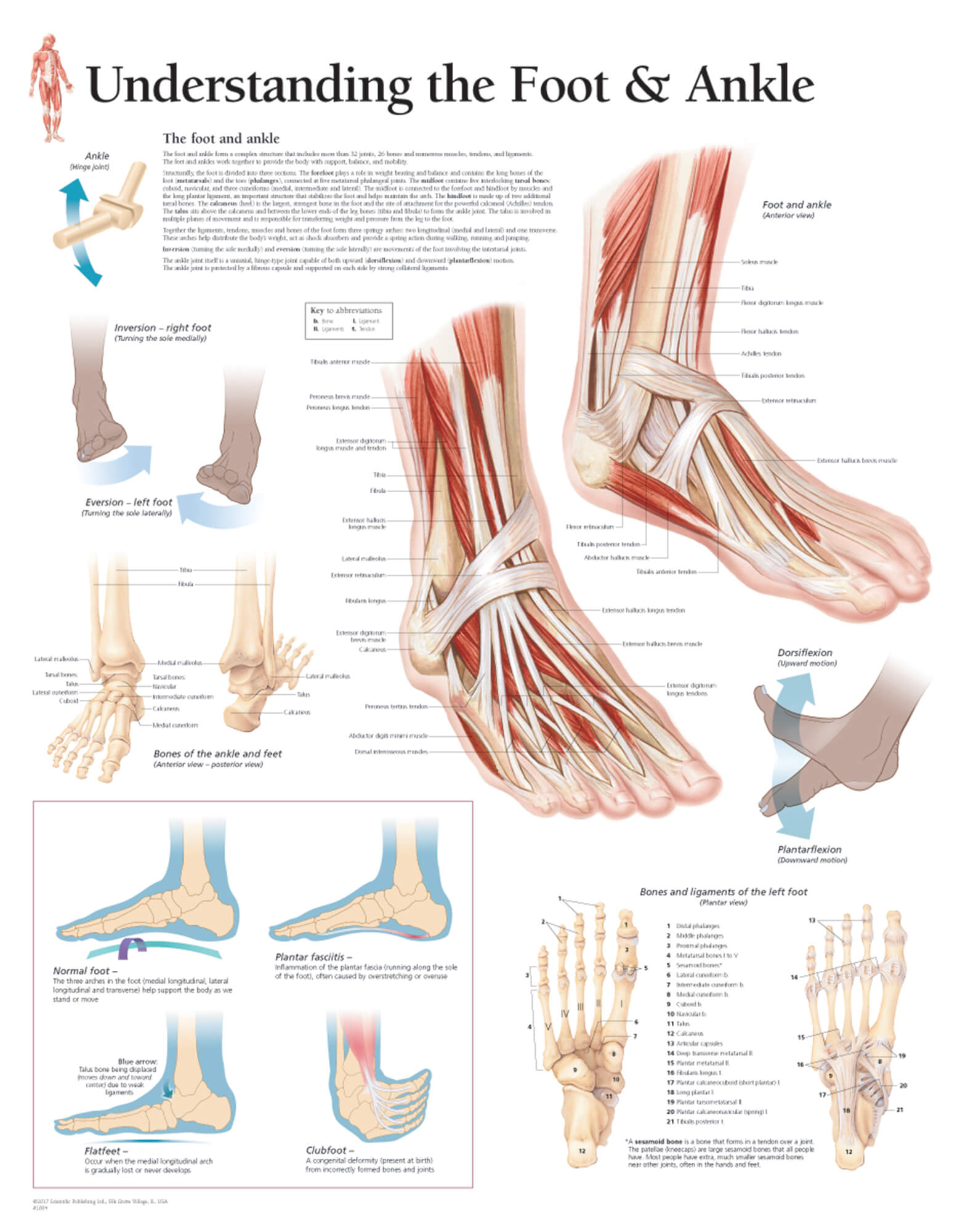
Understanding the Foot & Ankle Scientific Publishing
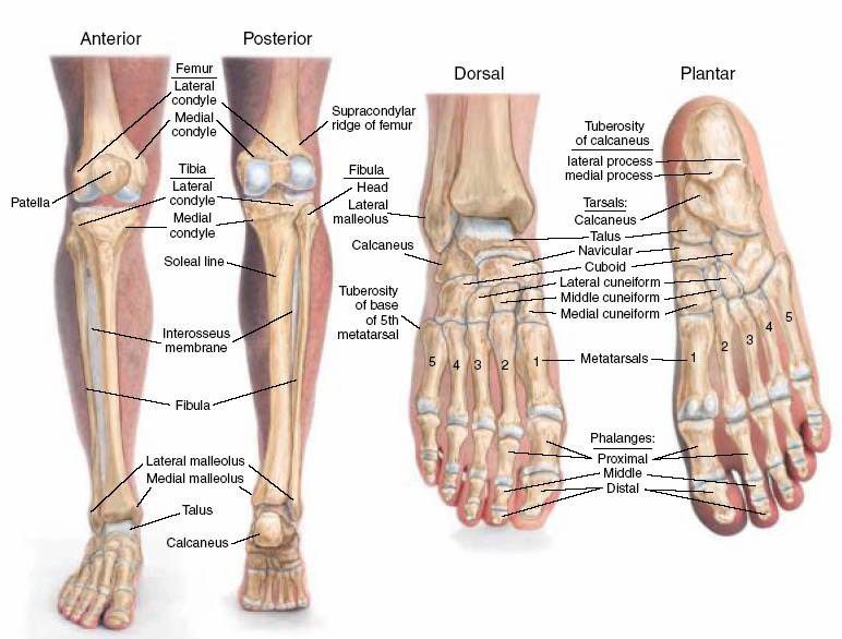
These bones are arranged in two rows, proximal and distal. The bones in the proximal row form the hindfoot, while those in the distal row from the midfoot. Hindfoot. Talus. Calcaneus. The talus connects the foot to the rest of the leg and body through articulations with the tibia and fibula, the two long bones in the lower leg. Midfoot. Navicular.
Human foot anatomy hires stock photography and images Alamy
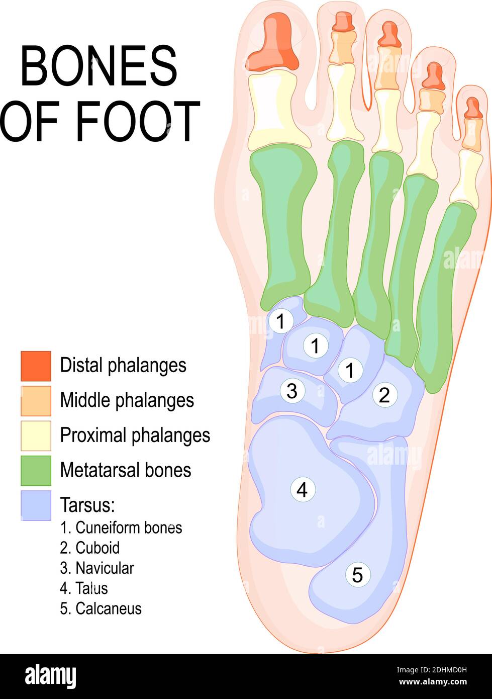
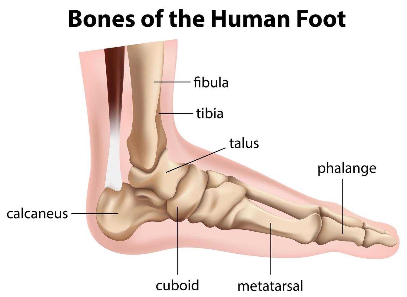
Bones of foot. The 26 bones of the foot consist of eight distinct types, including the tarsals, metatarsals, phalanges, cuneiforms, talus, navicular, and cuboid bones. The skeletal structure of.
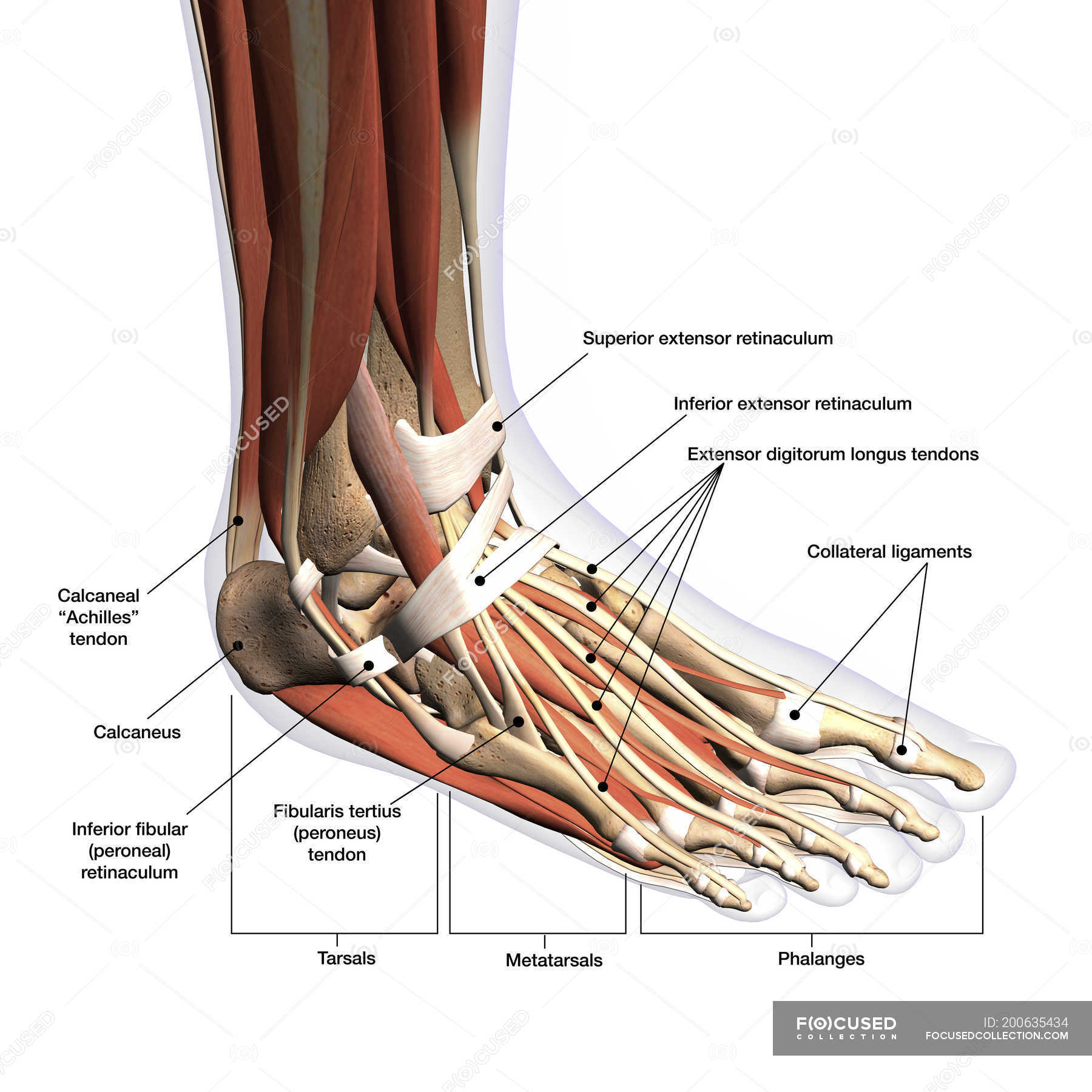
Anatomy of human foot with labels on white background — ankle, leg
Foot and ankle anatomy consists of 33 bones, 26 joints and over a hundred muscles, ligaments and tendons. This complex network of structures fit and work together to bear weight, allow movement and provide a stable base for us to stand and move on.

Chart of FOOT Dorsal view with parts name Vector image Stock Vector
The bones of the foot provide mechanical support for the soft tissues; helping the foot withstand the weight of the body whilst standing and in motion. They can be divided into three groups: Tarsals - a set of seven irregularly shaped bones. They are situated proximally in the foot in the ankle area. Metatarsals - connect the phalanges to.

Foot & Ankle Bones
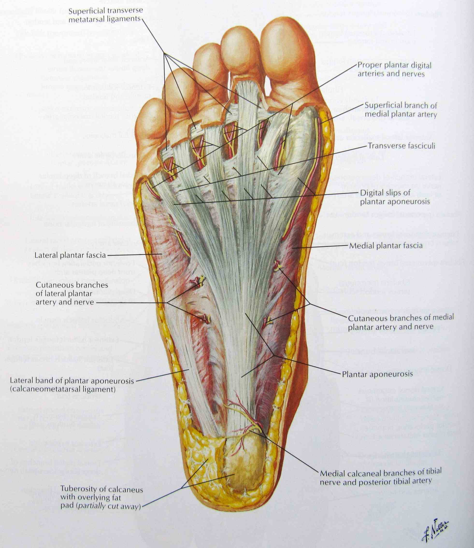
LABELED DIAGRAMS Figure 1. Sections and Bones of the Foot A. Lateral (Left) B. Anterior (Right) Figure 2. Compartments of the Foot A. Cut Section through Mid-Foot Figure 3. First Layer of the Foot A. Plantar View of Right Foot Figure 4. Second Layer of the Foot A. Plantar View of Right Foot Figure 5.

Anatomy of the Foot and Ankle OrthoPaedia
33 joints more than 100 muscles, tendons, and ligaments Bones of the foot The bones in the foot make up nearly 25% of the total bones in the body, and they help the foot withstand weight..
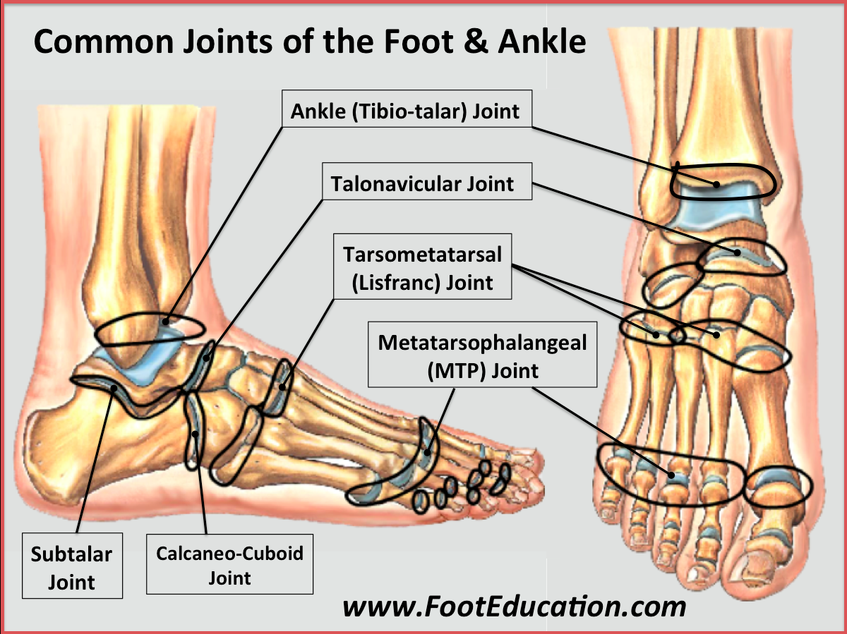
Bones and Joints of the Foot and Ankle Overview FootEducation
There are a variety of anatomical structures that make up the anatomy of the foot and ankle (Figure 1) including bones, joints, ligaments, muscles, tendons, and nerves. These will be reviewed in the sections of this chapter. Figure 1: Bones of the Foot and Ankle Regions of the Foot
.jpg)
Foot Bone Diagram resource Imageshare
The foot has three arches: two longitudinal (medial and lateral) arches and one anterior transverse arch (Fig. 1). They are formed by the tarsal and metatarsal bones, and supported by ligaments and tendons in the foot.
Anatomy The Bones Of The Foot
Plantar warts Gout (a type of arthritis) Plantar fasciitis (heel pain) Stress fractures Diabetic foot ulcers Last medically reviewed on April 13, 2015 The foot is the lowermost point of the.
Bones of the human foot diagram 1142236 Vector Art at Vecteezy
The foot is one of the most important interaction parts of the body with the ground, especially in the upright posture. During growth, the foot changes not only its dimensions but also its shape.

Foot Description, Drawings, Bones, & Facts Britannica
The anatomy of the foot The foot contains a lot of moving parts - 26 bones, 33 joints and over 100 ligaments. The foot is divided into three sections - the forefoot, the midfoot and the hindfoot. The forefoot This consists of five long bones (metatarsal bones) and five shorter bones that form the base of the toes (phalanges).

Diagram of The Foot 101 Diagrams
The 20-plus muscles in the foot help enable movement, while also giving the foot its shape. Like the fingers, the toes have flexor and extensor muscles that power their movement and play a large.

The bones in the foot inferior view (Picture illustrated from Thieme
The foot is the region of the body distal to the leg that is involved in weight bearing and locomotion. It consists of 28 bones, which can be divided functionally into three groups, referred to as the tarsus, metatarsus and phalanges. The foot is not only complicated in terms of the number and structure of bones, but also in terms of its joints.
Structure of skeleton of the foot, Tarsals, Metatarsals and Phalanges
When to see a doctor Summary The foot is an intricate part of the body, consisting of 26 bones, 33 joints, 107 ligaments, and 19 muscles. Scientists group the bones of the foot into the.

Foot bones Anatomy, conditions, and more
Common causes of foot pain include plantar fasciitis, bunions, flat feet, heel spurs, mallet toe, metatarsalgia, claw toe, and Morton's neuroma. If your feet hurt, there are effective ways to ease the pain. Some conditions specific to the foot can cause pain, less movement, or instability. Verywell / Alexandra Gordon.